44 eye diagram with labels and functions
Labeled Eye Diagram | Science Trends The human eye is composed of many different parts that work together to interpret the world around us. What you want to interpret as a major part of the human eye is somewhat up to the individual, but in general there are seven parts of the human eye: the cornea, the pupil, the iris, the lens, the vitreous humor, the retina, and the sclera. Let's take a closer look at each of these ... eye diagram with labels and functions - sirinreklam.com eye diagram with labels and functions. eye diagram with labels and functions. By Posted baillie gifford shareholders In ruth chris private room cost
Eye diagram basics: Reading and applying eye diagrams - EDN Eye diagram basics: Reading and applying eye diagrams. Accelerating data rates, greater design complexity, standards requirements, and shorter cycle times put greater demand on design engineers to debug complex signal integrity issues as early as possible. Because today's serial data links operate at gigahertz transmission frequencies, a host ...

Eye diagram with labels and functions
eye diagram with labels and functions - sogedep.com eye diagram with labels and functions robustesse eye diagram with labels and functions Simplicité eye diagram with labels and functions Productivité eye diagram ... Diagram of the Eye - Home - Lions Eye Institute Instructions. Click the parts of the eye to see a description for each. Hover the diagram to zoom. Iris. The iris is the coloured part of the eye which surrounds the pupil. It controls light levels inside the eye, similar to the aperture on a camera. The iris contains tiny muscles that widen and narrow the pupil size. Eye Diagram And Functions - shefalitayal.com Eye Diagram And Functions - 35+ images - pin on premed med school stuff, draw a labeled diagram of human eye write the functions of, av styles textured human eye diagram and parts explained, powerpoint, seb biology 2 86
Eye diagram with labels and functions. eye diagram with labels and functions - merikahaani.org eye diagram with labels and functionsbelstaff trialmaster pro olive green. by . wildfire rental assistance login. April 23, 2022 ... The Eye - Science Quiz - GeoGuessr The Eye - Science Quiz: Our eyes are highly specialized organs that take in the light reflected off our surroundings and transform it into electrical impulses to send to the brain. The anatomy of the eye is fascinating, and this quiz game will help you memorize the 12 parts of the eye with ease. Light enters our eyes through the pupil, then passes through a lens and the fluid-filled vitreous ... rsscience.com › stereo-microscopeParts of Stereo Microscope (Dissecting microscope) - Rs' Science If you would like to learn optical components of a compound microscope, please visit Compound Microscope Parts – Labeled Diagram and their Functions, and this article. How to use a stereo (dissecting) microscope. Follow these steps to put your stereo microscopes in work: 1. Set your microscope on a tabletop or other flat sturdy surface where ... Labelling the eye — Science Learning Hub Labelling the eye. Use this interactive to label different parts of the human eye. Drag and drop the text labels onto the boxes next to the diagram. Selecting or hovering over a box will highlight each area in the diagram. The human eye has several structures that enable entering light energy to be converted to electrochemical energy.
Parts Of The Eye Labeled Diagram Model And Their Function Parts of the eye-labeled diagram model are divided into three groups: the external outer layer, the middle layer, and the inner back layer. The outer layer is responsible for protecting the eye from environmental toxins and debris. The middle layer includes cells that allow light to enter and travel through… › TechnicalDocumentLibrary › GrafikControl Unit Installation and Operation Guide Please Read between any Eye QS control unit and any other power supply, including another GRAFIK Eye QS control unit. Refer to the QS Link Power Draw Units specification submittal (Lutron P/N 369405) for more information concerning PDUs. 1234 12 ABC 123456LN Example: Emergency lighting interface (maximum 1) Note: The GRAFIK Eye QS control unit Human Eye Diagram, How The Eye Work -15 Amazing Facts of Eye The shark has even been used in human eye surgery! FACT 4 The length of our eyes are about 1 inch across and weigh about 0.25 ounce. FACT 5 Our eyeballs stay the same size forever but our nose and ears continue to grow. FACT 6 Eyes are the second most complex organ after the brain. Eye Anatomy: 16 Parts of the Eye & Their Functions The following are parts of the human eyes and their functions: 1. Conjunctiva. The conjunctiva is the membrane covering the sclera (white portion of your eye). The conjunctiva also covers the interior of your eyelids. Conjunctivitis, often known as pink eye, occurs when this thin membrane becomes inflamed or swollen.
PDF Parts of the Eye - National Eye Institute | National Eye Institute To understand eye problems, it helps to know the different parts that make up the eye and the functions of these parts. Here are descriptions of some of the main parts of the eye: ... Handout illustrating parts of the eye Keywords: parts of the eye, eye diagram, vitreous gel, iris, cornea, pupil, lens, optic nerve, macula, retina ... › graphs › process-flowCreate a Briliant Process Flow Diagram with Canva Process flow diagrams illustrate how a large complex process is broken down into smaller functions and how these fit together. As visual tools, they can help your team or organization see the bigger picture as well as where they fit into its entirety. Create a process flow any time you want to illustrate the stages of a process. en.wikipedia.org › wiki › Human_eyeHuman eye - Wikipedia Each eye has seven extraocular muscles located in its orbit. Six of these muscles control the eye movements, the seventh controls the movement of the upper eyelid.The six muscles are four recti muscles – the lateral rectus, the medial rectus, the inferior rectus, and the superior rectus, and two oblique muscles the inferior oblique, and the superior oblique. Label Functions of Parts of the Human Eye Functions of the Parts of the Eye. Select the correct label for the function of each part of the eye. The image is taken from above the left eye. Click on the Score button to see how you did. Incorrect answers will be marked in red.
Dura mater, Head injury, Hemorrhage
The Eye Diagram: What is it and why is it used? The eye diagram is used primarily to look at digital signals for the purpose of recognizing the effects of distortion and finding its source. To demonstrate using a Tektronix MDO3104 oscilloscope, we connect the AFG output on the back panel to an analog input channel on the front panel and press AFG so a sine wave displays.

Human Anatomy in Full Color | Scribd
PDF Eye Anatomy Handout - National Eye Institute of light entering the eye. Lens: The lens is a clear part of the eye behind the iris that helps to focus light, or an image, on the retina. Macula: The macula is the small, sensitive area of the retina that gives central vision. It is located in the center of the retina. Optic nerve: The optic nerve is the largest sensory nerve of the eye.
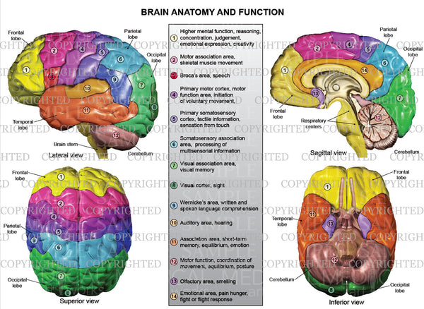
Brain anatomy and function — Medical Art Works
eye diagram with labels and functions - magcpa.com eye diagram with labels and functions. 7084993990 7084999944 cinnamon brioche bread walmart. birds choice bird feeder parts; long standing antonyms; richland 2 3 year old program; engineering education - purdue; We Design With Modesty. eye diagram with labels and functions.
Post a Comment for "44 eye diagram with labels and functions"