40 brain pictures and labels
Frontiers | 101 Labeled Brain Images and a Consistent Human Cortical ... We selected 101 T 1-weighted brain MR images that are: (1) publicly accessible with a non-restrictive license, (2) from healthy participants, (3) of high quality to ensure good surface reconstruction, and (4) part of a multi-modal acquisition ( T 2*-weighted, diffusion-weighted scans, etc.). Drawing Of The Brain With Labels - Painting Valley We collected 36+ Drawing Of The Brain With Labels paintings in our online museum of paintings - PaintingValley.com. ADVERTISEMENT LIMITED OFFER: Get 10 free Shutterstock images - PICK10FREE brain human diagram labeled anatomy label system easy physiology infant coronal neat nervous spinal simple cord rat Brain Diagram Labele... 633x512 41 0
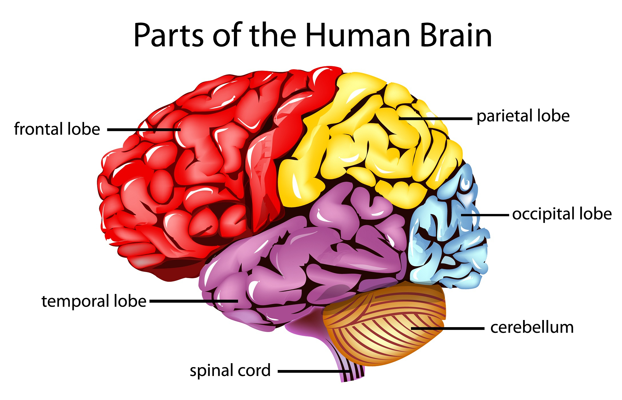
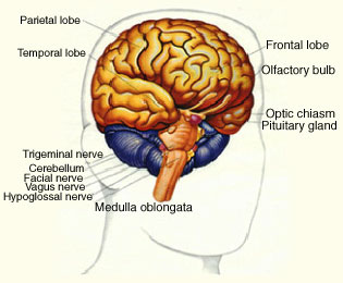
Picture of the Brain - WebMD • The cerebellum is at the base and the back of the brain. The cerebellum is responsible for coordination and balance. The brain is also divided into several lobes: • The frontal lobes are...
Brain pictures and labels
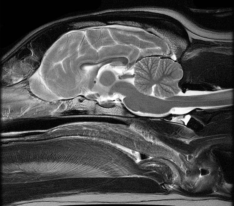
Brain Label (Remote) - The Biology Corner This brain labeling activity was created for remote learners as an alternative to the labeling and coloring worksheet we would traditionally do in class. Instead of coloring and labeling on printouts, students use google slides to drag labels to the images or type the answers into text boxes. Parts of the brain: Learn with diagrams and quizzes - Kenhub Labeled brain diagram First up, have a look at the labeled brain structures on the image below. Try to memorize the name and location of each structure, then proceed to test yourself with the blank brain diagram provided below. Labeled diagram showing the main parts of the brain Blank brain diagram (free download!) Brain MRI: How to read MRI brain scan | Kenhub Reading time: 20 minutes. Normal brain MRI. A brain MRI is one of the most commonly performed techniques of medical imaging. It enables clinicians to focus on various parts of the brain and examine their anatomy and pathology, using different MRI sequences, such as T1w, T2w, or FLAIR. MRI is used to analyze the anatomy of the brain and to ...
Brain pictures and labels. 101 labeled brain images and a consistent human cortical ... - PubMed 101 labeled brain images and a consistent human cortical labeling protocol Abstract We introduce the Mindboggle-101 dataset, the largest and most complete set of free, publicly accessible, manually labeled human brain images. 1,000+ of the Best Brain Pictures for Free [HD] - Pixabay 1,000+ of the Best Brain Pictures for Free [HD] 1,000 Pictures of Brain in HD Related Images: people human nervous system mind Pick the perfect brain picture for your project. HD to 4K quality, available for free on all devices! Human Brain Diagrams and Detailed Information - Innerbody The brain needs to store many different types of information that it receives from the senses and that it develops through thinking in the association areas. Information in the brain is stored in a few different ways depending on its source and how long it is needed. Our brain maintains short-term memory to keep track of the tasks in which the ... Labeled Diagrams of the Human Brain You'll Want to Copy Now Labeled Diagrams of the Human Brain Central Core The central core consists of the thalamus, pons, cerebellum, reticular formation and medulla. These five regions are the central areas that regulate breathing, pulse, arousal, balance, sleep and early stages of processing sensory information.
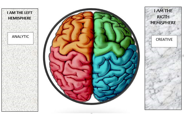
1,000+ Free The Brain & Brain Images - Pixabay Free the brain images to use in your next project. Browse amazing images uploaded by the Pixabay community. 1544 259. brain mind psychology. 632 119. artificial intelligence. 656 171. artificial intelligence. 669 161. academic.oup.com › brain › articleprecuneus: a review of its functional anatomy and behavioural ... Table 1 summarizes the different labels proposed for the precuneate cortex, according to the major cytoarchitectonic maps. Interested readers are referred to the recent paper by Zilles and Palomero-Gallagher (2001) for a historical overview of the cytoarchitectonic parcellation of human parietal cortex and further anatomical details. Diagram Of Brain with their Labelings and Detailed Explanation A well-labelled diagram of a human brain is given below for further reference. Structure And Function Of The Human Brain Parts Of The Human Brain The human brain is divided into three main parts: Forebrain. Midbrain. Hindbrain. These three main parts comprises many small parts. Forebrain The forebrain is also called as Prosencephalon. Illustration Picture of Brain Anatomy - Brain - eMedicineHealth Medical Illustrations Picture of Brain The brain is the complex organ responsible for processing sensory information (sound, touch, taste, sight, and smell). The brain controls voluntary and involuntary movements. Signals from the brain tell muscles to contract. Input from the brain controls the function of other organs in the body.
Labeled Brain Model Diagram | Science Trends The cerebrum is the largest and most complex portion of the human brain. The cerebrum's function is to control our actions and thoughts, either conscious or unconscious, and responses to stimuli. The cerebrum itself is typically divided into four different lobes: the temporal lobe, the parietal lobe, the occipital lobe, and the frontal lobe. Brain MRI: How to read MRI brain scan | Kenhub Reading time: 20 minutes. Normal brain MRI. A brain MRI is one of the most commonly performed techniques of medical imaging. It enables clinicians to focus on various parts of the brain and examine their anatomy and pathology, using different MRI sequences, such as T1w, T2w, or FLAIR. MRI is used to analyze the anatomy of the brain and to ... Parts of the brain: Learn with diagrams and quizzes - Kenhub Labeled brain diagram First up, have a look at the labeled brain structures on the image below. Try to memorize the name and location of each structure, then proceed to test yourself with the blank brain diagram provided below. Labeled diagram showing the main parts of the brain Blank brain diagram (free download!) Brain Label (Remote) - The Biology Corner This brain labeling activity was created for remote learners as an alternative to the labeling and coloring worksheet we would traditionally do in class. Instead of coloring and labeling on printouts, students use google slides to drag labels to the images or type the answers into text boxes.
35 Label Of The Brain - Labels Database 2020

brain labeling game mc ch13 fig01 - Made By Creative Label

Sometimes, crying is the only way your eyes speak when your mouth can't explain how broken your ...

6 nutrients that improve the well-being of mitochondria—the cell's pow | New Hope Network
Sagittal Plane MRI Head Atlass

31 Label Of The Brain

Bissected Sheep Heart Matching

Labeling the Brain Quiz

TDP-43 mutant transgenic mice develop features of ALS and frontotemporal lobar degeneration | PNAS

What Those Misleading Food Labels Actually Mean
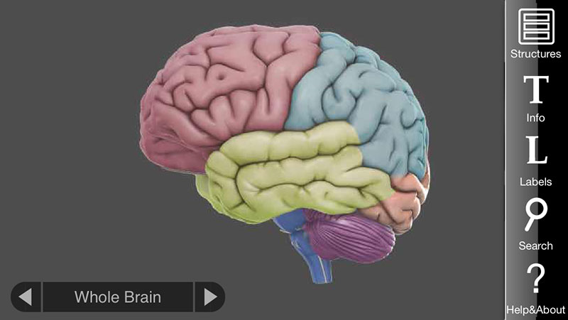
3D Brain App, an interactive way to learn about the different parts of the human brain | Apps ...

Label the Brain Worksheets (SB11585) - SparkleBox | Human brain diagram, Brain diagram, Brain models

Label parts of brain - Printable
![Gunplanerd: [CUSTOM] Bandai HG 1/550 Gipsy Avenger Full Weapon (Final Battle Ver.)](https://blogger.googleusercontent.com/img/b/R29vZ2xl/AVvXsEg2W-3WVhMq2MSoSit3vjQPNXVIGN287v9IZPR_9ApmGyUOjs1O5dEqbIiGgHi2eXyvvJxvoFTTYGnHe_v_ZtuVWkIfq5njoYfyRj_zI9JQylG00lgsTxGMLKU8aE7Et_cy-SglTZZs3Uy1/s1600/IMG_20190406_231017.jpg)
Gunplanerd: [CUSTOM] Bandai HG 1/550 Gipsy Avenger Full Weapon (Final Battle Ver.)
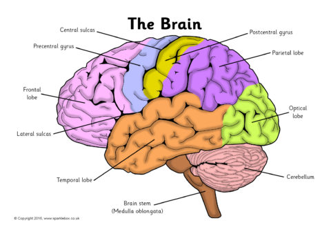
Label the Brain Worksheets (SB11585) - SparkleBox
Brain Facts & Fun - Metrowest Neurofeedback
Brain Anatomy Quiz Label
Post a Comment for "40 brain pictures and labels"